Electron Microscopy
Electron Microscopy Platform

Welcome
The Electron Microscopy Platform is an open access facility at the CIC bioGUNE. The mission of this platform is to offer high tech instrumentation, competitive service, specialized training and support in projects requiring transmission electron microscopy (TEM) to determinate the sub-nanometric structure of different types of samples.
This platform is mainly focused on structural biology for life science research. Nevertheless we also provide TEM analysis for material science users. Our platform offers research institutes and companies access to two cryo-transmission electron microscopes and sample preparation instrumentation. The sample preparation and data collection service is provided by the cryo-electron microscopy (cryo-EM) expert staff at the EM Platform.
- Platform Manager: Adriana L Rojas
Instrumentation
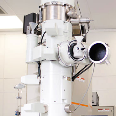
The core infrastructure of the EM Platform at CIC bioGUNE consists of two Jeol cryo transmission electron microscopes. They are flexible high contrast microscopes for the structural and morphological characterization of different types of samples. The most sophisticated microscope is the JEM-2200FS/CR, equipped with a 200-kV field emission gun (FEG) and an omega in-column energy filter. The other microscope is the JEM-1230, equipped with a 120-kV LaB6 thermionic gun.
Instrument list
- JEM-2200FS/CR transmission electron microscope (JEOL, Japan), equipped with an UltraScan 4000 SP (4008×4008 pixels) cooled slow-scan CCD camera (GATAN, UK)
- JEM-1230 transmission electron microscope (JEOL, Japan), equipped with an Orius SC1000 (4008×2672 pixels) cooled slow-scan CCD camera (GATAN, UK).
- Vitrobot, automated vitrification robot (FEI, The Netherlands).
- 3 cryotransfer holders used for cryo electron microscopy.
- 1 double tilt cryotransfer holder used for cryo electron microscopy.
- 1 ultra-high tilt cryotransfer holder used for cryo-electron tomography.
- Darkroom and Z/I PHOTOSCAN scanner (ZEIS).
- High vacuum coating system (MED 020 BALTEC) for carbon evaporation and glow discharge.
- HP Workstations with linux for data processing..

Service catalogue
Since there are many scientists that may be interested in incorporating EM studies in their own project, the EM platform at CIC bioGUNE offers the infrastructure and know-how as a specialized service, offering sample preparation, data collection and data processing for the specific user’s requirements, using single particle analysis or electron tomography techniques.
Service list
- Preparation for specific user requirements.
- TEM/Cryo-TEM data collection.
- Data processing and structure determination.
Specimen preparation techniques
There are many ways of preparing specimens for electron microscopy. In particular, a specimen preparation method for a biological sample perform the task of stabilizing the initially hydrated molecules, so that this sample can be placed and observed in the harsh vacuum of an electron microscope. Biological specimens at our laboratory are typically prepared using negative staining with heavy metal salts such as uranyl acetate producing high contrast, or by cryo-fixation techniques where the samples are embedded in vitreous ice in a frozen hydrated near native state.
Three-dimensional cryo-EM reconstruction of macromolecular assemblies
Cryo-transmission electron microscopy (cryo-TEM) is a low-dose imaging technique to visualize macromolecular structures, such as virus, proteins and pieces of cell organelles, in a near native state with sub-nm resolution. Depending on the specific question and or sample, two different cryo-EM data processing methodologies can be used to form an accurate three-dimensional (3D) representation (3D-map) of the biological object with sub-nm resolution, from a set of 2D noisy cryo-EM images. One of these methods is Single Particle Analysis and another method is Cryo-Electrom Tomography (cryo-ET). On the one hand, for macromolecular assemblies (in the size range of ~5-50 nm) that have identical structure by functional necessity, powerful averaging techniques can be used to eliminate noise and obtain near atomic resolution 3D maps (example. Virus, ribosomes …). On the other hand, for cell components (in the size range of ~100-1000 nm), which possess a unique structure, (example. Mitochondrion, bacteria …), they are visualized as “individuals” by obtaining one entire projection series that is aligned and reconstructed to produce a tomogram.
Service Request
For inquiries and consultations on Electron Microscopy (Cryo-EM, Cryo-ET, Cryo-TEM) projects, please contact directly with Adriana L Rojas emicroscopyplatform@cicbiogune.es


